
The pervasive infiltration of microscopic plastic particles into Earth’s ecosystems and biological organisms represents a pressing global concern. While these ubiquitous fragments, spanning both microplastics and their even smaller counterparts, nanoplastics, have been definitively identified across diverse environments—from the abyssal depths of oceans to fertile agricultural lands and the intricate tissues of wildlife and humans—a significant void persists in our comprehensive understanding of their precise journey and transformations once assimilated into living systems. A recent scientific breakthrough introduces an innovative fluorescence-based methodology, poised to revolutionize the real-time observation of microplastic transit, chemical evolution, and ultimate degradation within biological matrices.
The Ubiquity of Plastic Contamination and Its Enigma
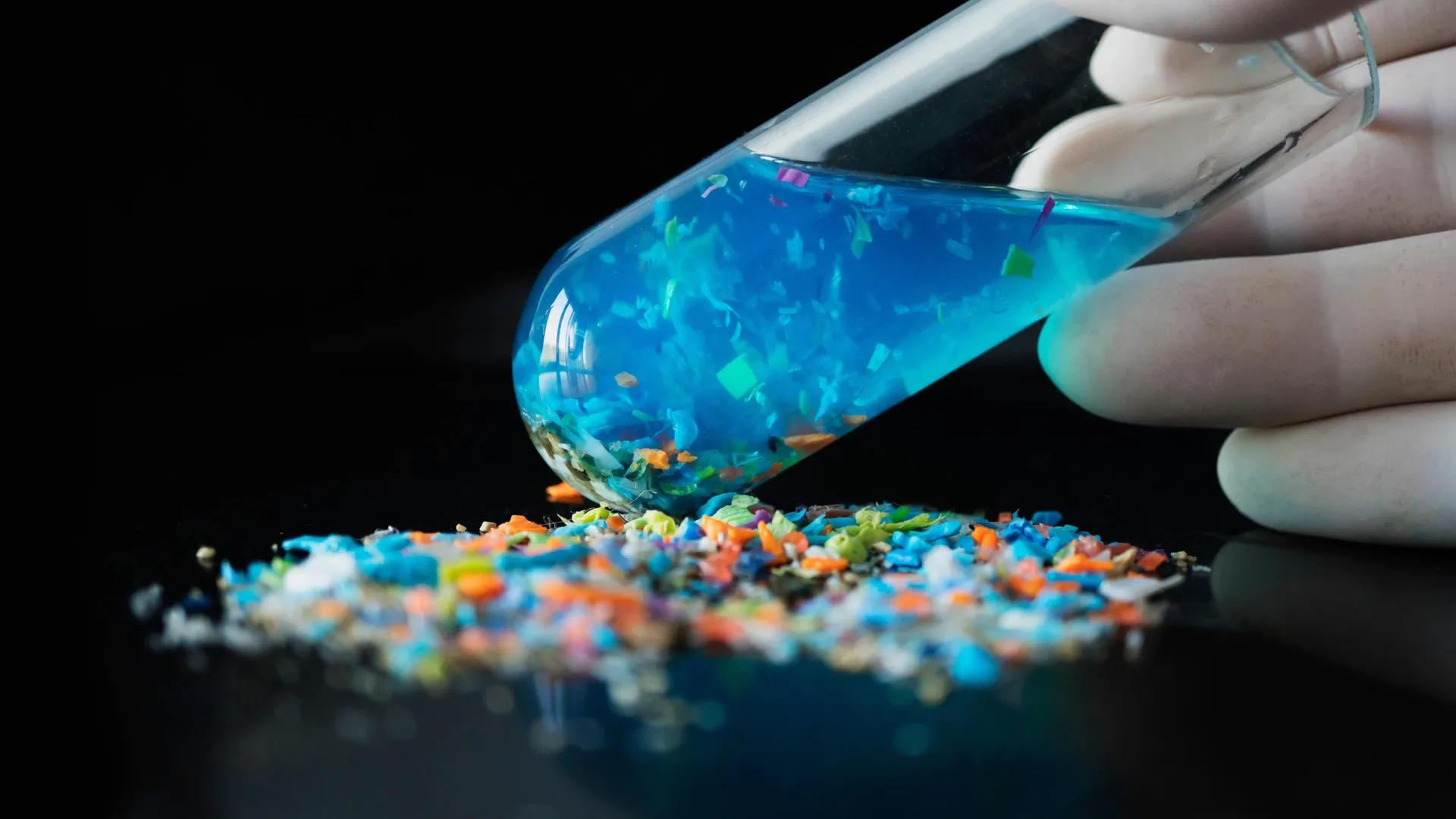
Global plastic production has surged past 460 million metric tons annually, contributing millions of tons of microscopic plastic debris into the environment each year. This persistent anthropogenic material has been detected in a startling array of biological samples, including marine fauna, avian species, and a growing list of human tissues such as blood plasma, hepatic cells, and even cerebral matter. While preliminary laboratory investigations have suggested potential links between microplastic exposure and adverse health outcomes, including inflammatory responses, cellular damage, and developmental aberrations, the fundamental mechanisms governing their interaction with biological systems remain largely undefined. The precise routes of entry, patterns of distribution, rates of accumulation, and ultimate fate within a living organism constitute a critical knowledge gap that impedes robust risk assessment and the formulation of effective mitigation strategies.
The journey of plastics from consumer products to environmental contaminants and ultimately into biological systems is complex and multifaceted. Primary microplastics are intentionally manufactured as small beads or fibers for industrial or cosmetic applications. Secondary microplastics, far more prevalent, arise from the fragmentation of larger plastic items dueoplastics degrade under environmental stressors like UV radiation, mechanical abrasion, and biological activity. These fragments, ranging from micrometers to millimeters in size, possess diverse shapes—fibers, fragments, spheres, and films—and varying chemical compositions, depending on the polymer type (e.g., polyethylene, polypropylene, polystyrene, PVC, PET). Nanoplastics, typically defined as particles smaller than 100 nanometers, represent an even greater challenge due to their minuscule dimensions, which theoretically allow for easier translocation across biological barriers and into cellular structures. Their omnipresence in air, water, and soil ensures constant exposure pathways for humans and wildlife through inhalation, ingestion of contaminated food and water, and dermal contact.
Limitations of Conventional Analytical Paradigms
Current analytical techniques employed for detecting and characterizing microplastics in biological samples predominantly suffer from inherent limitations that preclude dynamic, real-time observation. Spectroscopic methods, such as Fourier-transform infrared (FTIR) spectroscopy and Raman spectroscopy, provide detailed chemical fingerprints of plastic polymers but typically necessitate sample preparation steps that can be destructive, involving the digestion or incineration of tissues to isolate the plastic components. Similarly, mass spectrometry techniques, while offering high sensitivity and specificity for polymer identification, also require sample destruction. This destructive nature means that researchers can only obtain static "snapshots" of microplastic presence at specific time points, preventing the continuous monitoring of their movement, chemical alteration, or breakdown within an intact, living organism. This inability to track the spatiotemporal dynamics within a complex biological milieu has severely hampered efforts to elucidate critical questions, such as: What are the primary uptake mechanisms? How do microplastics traverse cellular and tissue barriers? What is their residence time in different organs? Do they bioaccumulate or biodegrade, and into what byproducts? And how do their physical and chemical properties evolve over time in a biological environment?
Fluorescence imaging, an established technique in biological research, presents a promising avenue for non-invasive, real-time tracking. However, its application to microplastics has historically been fraught with challenges. Traditional methods often involve coating plastic particles with external fluorescent dyes. These dyes are susceptible to photobleaching, where their luminescence diminishes over time due to light exposure. Furthermore, the dyes can leach from the plastic surface into the surrounding biological environment, leading to false signals or reduced signal intensity. The complex and often autofluorescent nature of biological tissues can also quench the signal from external dyes, further diminishing their effectiveness in providing clear, stable, and persistent tracking. These issues underscore the need for a more robust and integrated fluorescent labeling strategy.
Pioneering a Novel Fluorescent Tracking Modality
To surmount these significant methodological hurdles, a research team has engineered an innovative strategy termed "fluorescent monomer controlled synthesis." This groundbreaking approach fundamentally deviates from conventional surface-coating techniques. Instead of merely affixing fluorescent dyes to the exterior of plastic particles, the light-emitting components are meticulously incorporated directly into the polymer’s molecular architecture during the plastic synthesis process itself. This intrinsic integration ensures that the fluorescent markers are an integral part of the plastic material, not merely an external addition.
A cornerstone of this advanced methodology lies in the utilization of Aggregation-Induced Emission (AIE) materials. Unlike traditional fluorophores, which often experience a phenomenon known as aggregation-caused quenching (ACQ)—where their luminescence dramatically diminishes when they are clustered together—AIE materials exhibit the inverse behavior. They remain largely non-emissive or weakly fluorescent when dispersed as individual molecules but become intensely luminescent when aggregated or confined. This characteristic is particularly advantageous in biological systems, where particles are often densely packed or accumulate within cells and tissues. The AIE mechanism ensures a stable and robust signal, significantly mitigating issues of photobleaching and dye leakage that plague conventional fluorescent labels. By embedding these AIE components directly into the polymer matrix, the method ensures a persistent and brightly emissive signal throughout the particle’s existence.
This sophisticated polymer engineering approach offers unparalleled versatility and control over the properties of the microplastic tracers. Researchers can precisely fine-tune several key parameters, including:
- Brightness: Adjusting the concentration and type of AIE luminogens allows for tailored signal intensity, optimizing visibility for diverse biological contexts.
- Emitted Light Color: The spectral properties of the AIE materials can be modulated to produce different emission wavelengths, enabling multi-color tracking or differentiation of various plastic types simultaneously.
- Particle Size and Shape: The synthesis process allows for the creation of fluorescent microplastics with defined dimensions and morphologies, enabling investigations into how these physical characteristics influence biological interactions.
Crucially, because the fluorescent material is homogeneously distributed throughout the entire structure of each synthesized plastic particle, its visibility is maintained not only when the particle is intact but also as it undergoes degradation. This capability means that even the smaller fragments generated during the breakdown process remain fluorescent and trackable. This unprecedented feature opens the door to monitoring the complete lifecycle of microplastics within an organism, encompassing initial ingestion, internal translocation across biological barriers, subsequent chemical transformation and physical fragmentation, and ultimately, their final breakdown products and clearance pathways.
Transformative Implications for Health and Environmental Risk Assessment
While this innovative strategy is currently undergoing rigorous experimental validation, its foundation rests upon well-established principles derived from polymer chemistry and biocompatible fluorescence imaging. The scientific community anticipates that this approach will evolve into an indispensable tool for deciphering the intricate interactions between microplastics and fundamental biological entities, including individual cells, complex tissues, and whole organ systems.
The capacity for dynamic, real-time tracking of microplastics represents a paradigm shift in environmental health research. By providing continuous insights into the transport and transformation processes within living organisms, this technology will enable a more nuanced and accurate assessment of the true ecological and health risks posed by plastic pollution. This moves beyond merely quantifying exposure levels to understanding the precise mechanisms of toxicity. For instance, researchers will be able to investigate whether specific plastic types or sizes preferentially accumulate in certain organs, whether their surface chemistry changes in response to biological fluids, and how these alterations might influence their toxicological profile. This depth of understanding is vital for establishing robust dose-response relationships and identifying particularly vulnerable populations or physiological systems.
The implications extend far beyond basic research. Enhanced understanding of microplastic dynamics could directly inform public health guidelines, refine environmental regulations, and stimulate the development of targeted intervention strategies. For instance, if certain plastic formulations are found to degrade into more toxic byproducts within the body, this could lead to calls for their restriction or replacement. Furthermore, this technique could be adapted for screening the biocompatibility of new materials, assessing drug delivery systems that utilize similar polymer carriers, and even in fundamental cell biology studies exploring particle uptake and trafficking.
As global apprehension regarding the pervasive nature of plastic pollution intensifies, the development of sophisticated analytical tools capable of illuminating the complex behavior of microplastics within living systems is paramount. This fluorescent tracking technology represents a significant leap forward, offering the potential to transform our understanding of plastic-biology interactions, thereby playing a pivotal role in refining risk assessments, guiding future policy decisions, and ultimately safeguarding both human health and planetary ecosystems. The ability to visualize the microscopic odyssey of plastic particles promises to unlock critical insights needed to confront one of the defining environmental challenges of our era.