
 Ayu Aulia
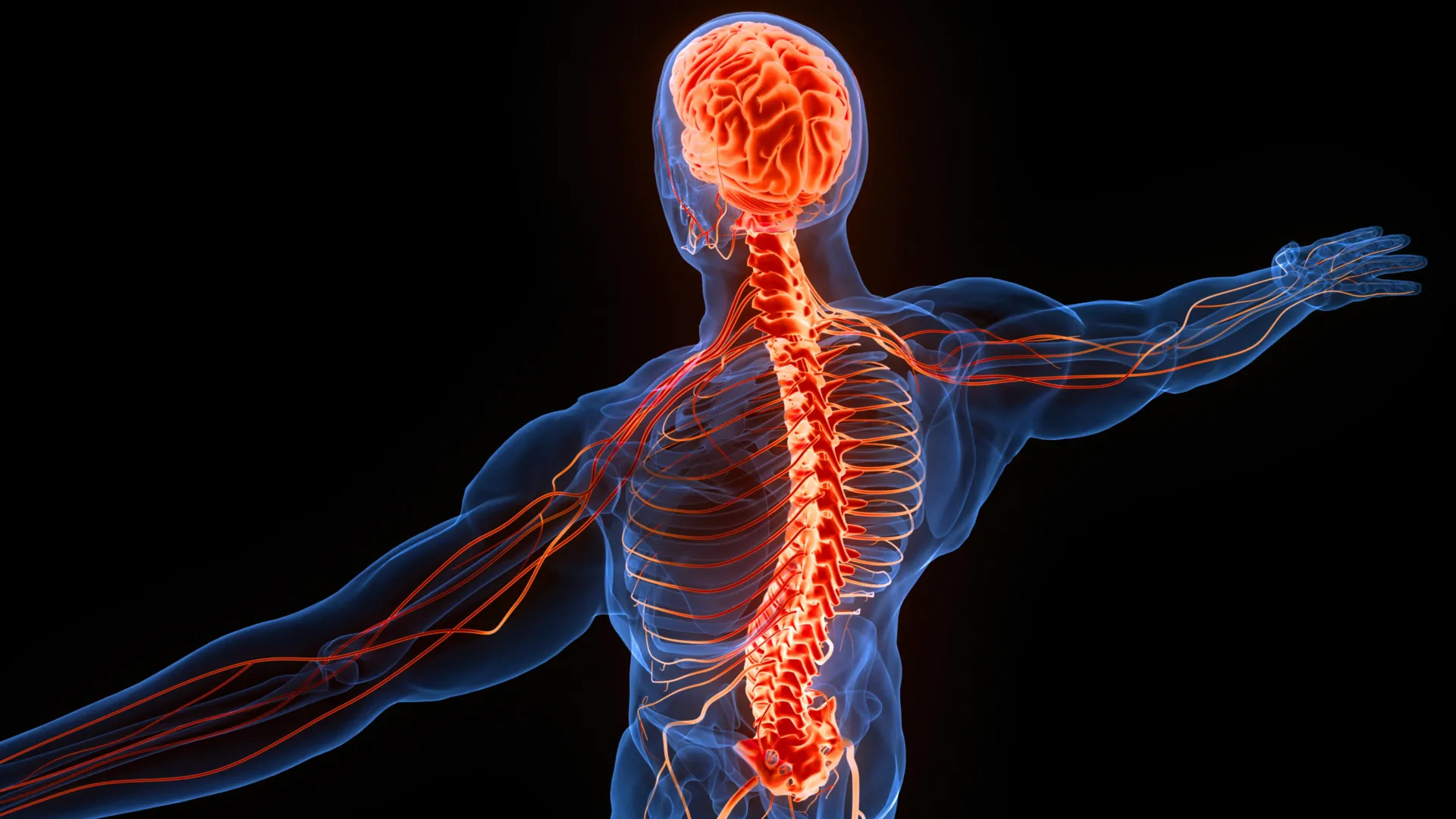
The peculiar phenomenon of neural decussation, where each cerebral hemisphere governs the opposite side of the body, has long presented a fundamental puzzle in neuroscience. This ubiquitous anatomical arrangement, a a defining characteristic of the vertebrate nervous system, is now being re-evaluated through a novel evolutionary lens. Recent research suggests that this intricate cross-wiring may not primarily be a directly advantageous adaptation for motor control or sensation, but rather an inherited organizational principle rooted in primordial visual processing that emerged hundreds of millions of years ago.
For centuries, the inverted relationship between brain hemispheres and bodily control has fascinated anatomists and neuroscientists. Why does the left brain manage the right side of the physique, and vice versa? Numerous hypotheses have been posited, ranging from theories of body torsion in early vertebrates to optimizing neural pathway lengths. However, a comprehensive explanation has remained elusive. A recent interdisciplinary examination, integrating insights from neuroanatomy, evolutionary biology, and comparative physiology, offers a compelling new framework. This perspective posits that the foundational architecture for this contralateral control was established by the earliest visual processing centers, which subsequently served as an immutable blueprint for the development of later sensory and motor pathways.
The Foundational Role of Visual Processing
The proposed explanation for neural decussation initiates with the fundamental mechanics of vision. When light traverses the lens of an eye, the resulting image projected onto the retina is inherently inverted. Consequently, objects situated in an organism’s right visual field are registered on the left half of each retina, while stimuli from the left visual field are mapped onto the right half. This optical inversion is a universal physical principle, independent of biological evolution.
Following retinal processing, visual information embarks on a complex journey through the nervous system. A critical juncture occurs at the optic chiasm, a structure where nerve fibers from the medial halves of the retinas cross over to the contralateral side of the brain. This crossing ensures that all visual data originating from the right visual field ultimately converges on the left cerebral hemisphere, and conversely, information from the left visual field is directed to the right hemisphere. This sophisticated re-organization is crucial for coherent visual perception and depth processing.
Further upstream, some of these reorganized visual signals project to an evolutionarily ancient brain structure known as the optic tectum. In mammals, this region is homologously referred to as the superior colliculus. Situated within the midbrain, the optic tectum plays a pivotal role in mediating rapid, reflexive responses to visual stimuli, such as orienting the head and eyes towards a sudden movement in the periphery. Each side of the optic tectum maintains a topographic representation of the opposite visual field. Intriguingly, deeper layers within this structure also integrate somatosensory information (physical sensations), organized into corresponding contralateral maps. The profound evolutionary conservation of the optic tectum, present in every vertebrate species, underscores its antiquity and suggests its critical importance as an organizational hub within the primordial nervous system. Its early establishment and inherently contralateral mapping of visual input are considered central to the new hypothesis.
Tracing the Contralateral Path: Sensory Information
The investigation extends beyond the visual system, meticulously tracing the evolutionary trajectory of major sensory pathways responsible for transmitting bodily sensations to the brain. Two prominent pathways, both exhibiting contralateral organization, are particularly scrutinized for their widespread presence among tetrapods—the four-limbed vertebrates encompassing mammals, birds, reptiles, and amphibians.
The first, termed the dorsal column-medial lemniscal pathway, is responsible for conveying highly discriminative tactile information, such as gentle touch, vibration, and proprioception (the sense of body position and movement). Signals originating from the limbs and torso ascend ipsilaterally (on the same side) through the dorsal columns of the spinal cord. Upon reaching specific nuclei within the brainstem, just superior to the spinal cord, these fibers synapse with second-order neurons. It is at this precise point within the brainstem that the pathway decussates, crossing to the contralateral side before continuing its ascent to the thalamus and ultimately the somatosensory cortex. While this pathway is absent in fish, its consistent presence across amphibians, reptiles, birds, and mammals strongly suggests its emergence early in tetrapod evolution and its subsequent remarkable conservation, highlighting its fundamental role in complex motor control and environmental interaction.
The second significant pathway, the spinothalamic tract (or anterolateral system), mediates the transmission of sensations crucial for survival, including pain, temperature, crude touch, and itch. Unlike the dorsal column pathway, the spinothalamic tract decussates much earlier in its trajectory, crossing to the opposite side within the spinal cord itself, typically within a few segments of its origin. After crossing, these signals ascend to the thalamus, a critical relay station in the midbrain for all sensory information (except olfaction), before projecting to the cerebral cortex. The contralateral organization of the spinothalamic tract is observed not only in birds, mammals, and reptiles but also in some cartilaginous fish, such as sharks and rays. This broader phylogenetic distribution implies an even deeper evolutionary lineage for this particular contralateral arrangement, hinting at its presence in very early vertebrate forms.
Controlling Movement: The Decussated Motor Landscape
The principle of contralateral organization is not confined to sensory processing; it extends profoundly into the control of skeletal muscles. The research identified three crucial motor pathways that similarly connect one side of the brain with the opposite side of the spinal cord, orchestrating movement across the body.
The first is the tectospinal pathway, a critical circuit for rapid, reflexive orienting movements of the head and neck in response to sensory input. As previously noted, the optic tectum (superior colliculus in mammals) is a multimodal sensory integration center. It processes visual information, but also receives and maps auditory and somatosensory inputs, aligning these signals spatially with its contralateral visual representation. For instance, if an individual is alerted by a sudden sound or a flash of light in their peripheral vision, the tectospinal pathway rapidly coordinates the reflexive turning of the head and eyes towards the stimulus. Movement signals originate in the deepest layers of the optic tectum, decussate, and descend through nerve fibers to innervate motor neurons on the opposite side of the spinal cord. This pathway’s prevalence across most tetrapods and certain cartilaginous fish reinforces its ancient origins and its fundamental role in mediating environmental engagement.
Another significant motor circuit is the rubrospinal pathway, believed to play a vital role in coordinating limb movements, particularly in locomotion and posture. Originating in the red nucleus, a distinct structure in the midbrain, this pathway also projects to the contralateral side of the spinal cord. While especially prominent in most mammals, contributing to arm and hand movements (though less finely than the corticospinal tract), its presence has also been documented in other tetrapods and some cartilaginous fish. This indicates a relatively ancient evolutionary development, predating the sophisticated motor control seen in higher primates.
The Evolution of Fine Motor Control: A Human Specialization
The third motor pathway, the corticospinal tract, holds particular significance for humans due to its indispensable role in mediating highly precise, voluntary movements, exemplified by the intricate dexterity of our fingers. Its evolutionary history, however, is notably more complex and exhibits greater species-specific variation compared to the other pathways.
While all vertebrates possess a telencephalon—the anterior-most part of the brain that constitutes a substantial portion of the human brain’s overall size—only mammals have evolved a cerebral cortex organized into six distinct layers. This neocortex is the substrate for sophisticated cognitive functions, including complex sensory integration, memory formation, motor planning, and language processing.
The corticospinal pathway originates within various regions of the cerebral cortex, primarily the motor cortex, and descends towards the spinal cord. Its functional emphasis, however, varies across species. In most non-primate mammals, the corticospinal tract primarily exerts an indirect influence, modulating sensory processing by projecting to spinal cord regions that receive afferent information from the body. This allows the brain to fine-tune and regulate incoming sensory signals.
In primates, including humans, and certain other animals renowned for their highly precise manipulative abilities, the corticospinal pathway has undergone significant evolutionary elaboration. Beyond its sensory modulatory role, it also establishes direct connections with motor neurons within the spinal cord that control specific muscles. These direct connections are the anatomical substrate for intricate, finely tuned actions, such as the coordinated finger movements required for tool use, writing, or playing a musical instrument. Crucially, regardless of its specific function (sensory modulation or direct motor control), the vast majority of the corticospinal tract’s fibers decussate, typically at the level of the brainstem in a prominent feature known as the pyramidal decussation.
This evolutionary trajectory suggests that the advanced fine motor capabilities unique to humans and other primates may have emerged not from the de novo creation of an entirely new neural system, but rather through the evolutionary co-option and functional adaptation of an older neural circuit. This pre-existing circuit, initially involved in regulating sensory input, gained an additional, highly specialized purpose: direct, voluntary motor control. This represents a remarkable example of evolutionary parsimony, where existing structures are repurposed and refined to meet new adaptive challenges.
A 500-Million-Year-Old Explanation: Evolutionary Inheritance
Synthesizing the evidence from these diverse neural pathways, a potentially profound explanation for the brain’s unusual contralateral organization emerges. The central hypothesis proposes that the earliest visual circuits, particularly the optic chiasm and optic tectum, played a pivotal role in establishing an initial, fundamental pattern of contralateral organization for sensory and movement pathways. Subsequent neural circuits, as they evolved and became more complex, may have largely inherited and conformed to this pre-existing architectural template.
Given that every extant vertebrate possesses an optic chiasm and an optic tectum—both organized around the principle of processing information from the opposite visual field—researchers infer that these structures were likely present in the primitive jawless fish that roamed the oceans during the Cambrian Period, approximately 538 to 486 million years ago. If this inference holds true, it implies that the fundamental organizational principle underlying the brain’s cross-connections began its development more than half a billion years before the emergence of modern humans.
This compelling hypothesis is not merely theoretical; it presents avenues for empirical validation. Future investigations could involve examining fossilized skulls of ancient jawless fish for anatomical evidence, such as specific neural canal structures or optic nerve pathways, that might corroborate the presence and organization of an optic chiasm and optic tectum in these earliest vertebrates. Advanced imaging techniques applied to well-preserved fossil specimens could provide crucial insights into the neuroanatomy of our most distant ancestors.
Evolutionary Inertia vs. Direct Advantage
The broader implications of this hypothesis challenge a pervasive assumption in evolutionary biology: that every unusual biological feature must have evolved because it conferred a direct and immediate survival advantage. While many adaptations undeniably offer such benefits, this new perspective suggests that the brain’s cross-wiring might be an enduring example of "evolutionary inertia" or "developmental constraint."
Evolution frequently operates by modifying existing structures and pathways rather than consistently constructing entirely novel systems from scratch. Once the ancient visual pathways established a contralaterally organized framework, this deeply embedded arrangement may have exerted a powerful influence on the subsequent development and wiring of additional sensory and motor circuits. In essence, the organization of the visual system, particularly the optic tectum, may have provided a robust structural foundation—a "default setting"—that later pathways found evolutionarily expedient to adopt.
Under this framework, the brain’s crossed connections did not necessarily arise because controlling the opposite side of the body was inherently more efficient or advantageous in a direct, functional sense. Instead, they may reflect a deep-seated historical legacy, a testament to an ancient organizational principle that evolution repeatedly reused and adapted over vast stretches of geological time. This perspective underscores how fundamental architectural decisions made in the earliest stages of vertebrate evolution could reverberate across millions of years, shaping the most complex aspects of modern neurobiology.
The result is one of the most profound and enduring examples of how the nervous system preserves its evolutionary history. The intricate and seemingly counterintuitive relationship between the brain’s two hemispheres and the opposite sides of the body may ultimately trace its origins back to the visual systems of pioneering vertebrate life forms that navigated ancient waters more than 500 million years ago, offering a remarkable window into the deep past of our own neural architecture. Further comparative neuroanatomical studies across diverse taxa, coupled with insights from developmental biology and genetic analysis, will continue to refine our understanding of this ancient and enduring enigma.




 Aura Kasih

 Lisa Mariana