A groundbreaking advancement in neural engineering has emerged from collaborative research, unveiling an extraordinarily compact brain implant capable of wirelessly transmitting intricate neural activity for over a year from living subjects, signifying a profound leap in our capacity to comprehend and interact with the nervous system.
For decades, the aspiration of scientists and medical professionals has been to develop seamless, long-duration interfaces with the brain. Understanding the brain’s complex circuitry, its responses to disease, and its mechanisms for learning and memory necessitates tools that can observe neural activity without disruption, for extended periods. Traditional brain-computer interfaces (BCIs), while making significant strides in areas like prosthetics control and communication for individuals with severe paralysis, have often been encumbered by limitations: the need for wired connections, larger physical footprints that risk tissue damage, finite operational lifespans, and challenges related to biocompatibility and chronic immune response. These constraints have historically hampered truly chronic, minimally invasive studies and clinical applications. The recent innovation, detailed in leading scientific publications, directly addresses many of these formidable challenges, heralding a new paradigm in neuro-monitoring and therapeutic intervention.
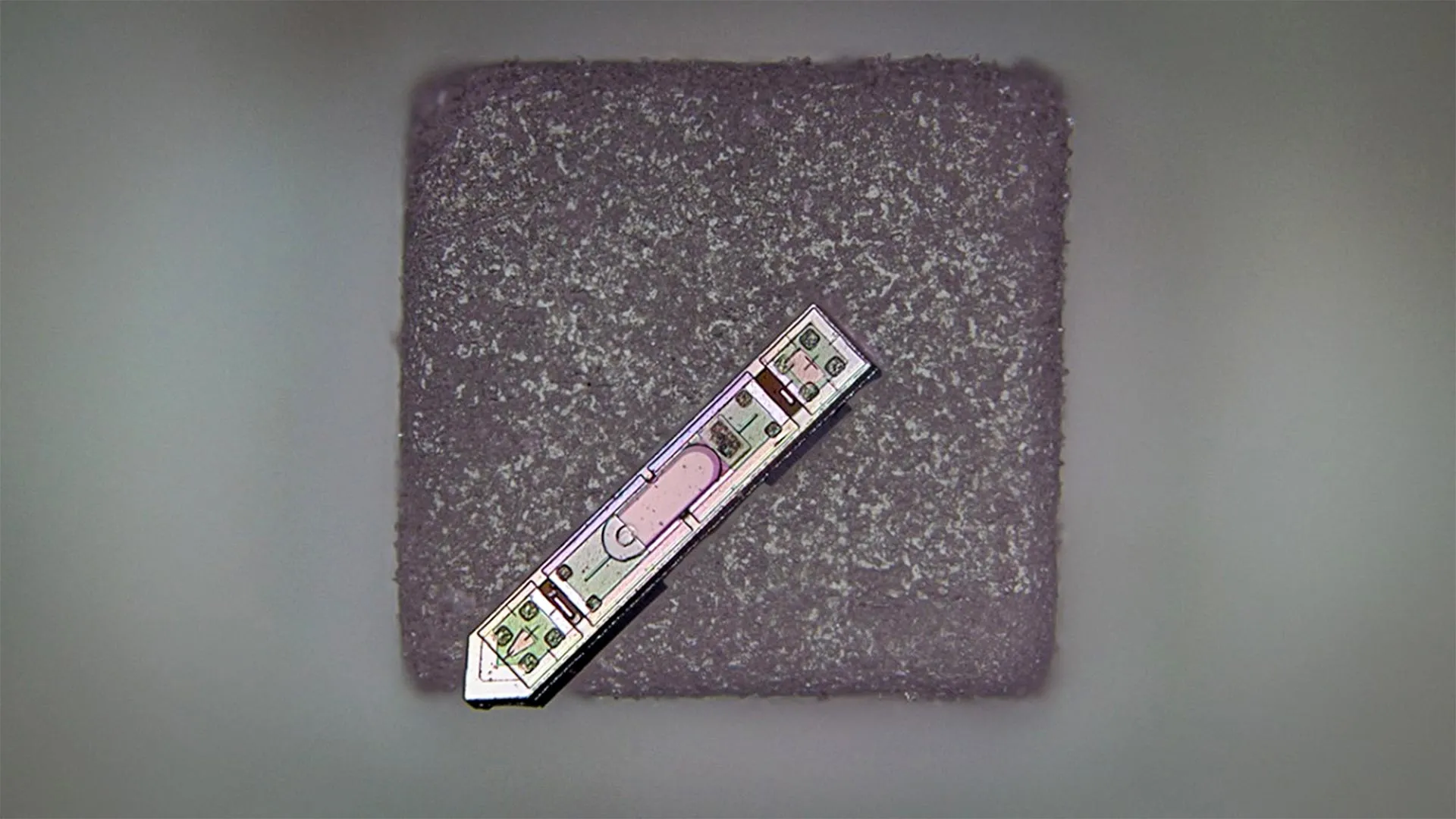
At the heart of this technological breakthrough is a device that pushes the boundaries of microelectronic miniaturization. Referred to by its creators as a microscale optoelectronic tetherless electrode, or MOTE, this implant represents a remarkable feat of engineering. Its dimensions are astonishingly small, measuring approximately 300 microns in length and 70 microns in width. To contextualize this, a human hair typically ranges from 17 to 180 microns in diameter, meaning this entire device is barely wider than an average human hair and shorter than the width of a single printed period on this page. Such an ultra-compact form factor is critical, as it drastically reduces the potential for tissue displacement and inflammatory responses within the delicate neural environment, a primary concern for any long-term implant. This minute scale also opens avenues for deploying multiple implants simultaneously, enabling high-density recording across broader brain regions, a capability that was previously difficult or impossible with larger, more intrusive devices. The engineering team meticulously designed the MOTE to be not only small but also robust enough to function reliably within the dynamic biological milieu of the brain, maintaining its structural and functional integrity over extended periods, an achievement that sets it apart from previous attempts at miniaturization.
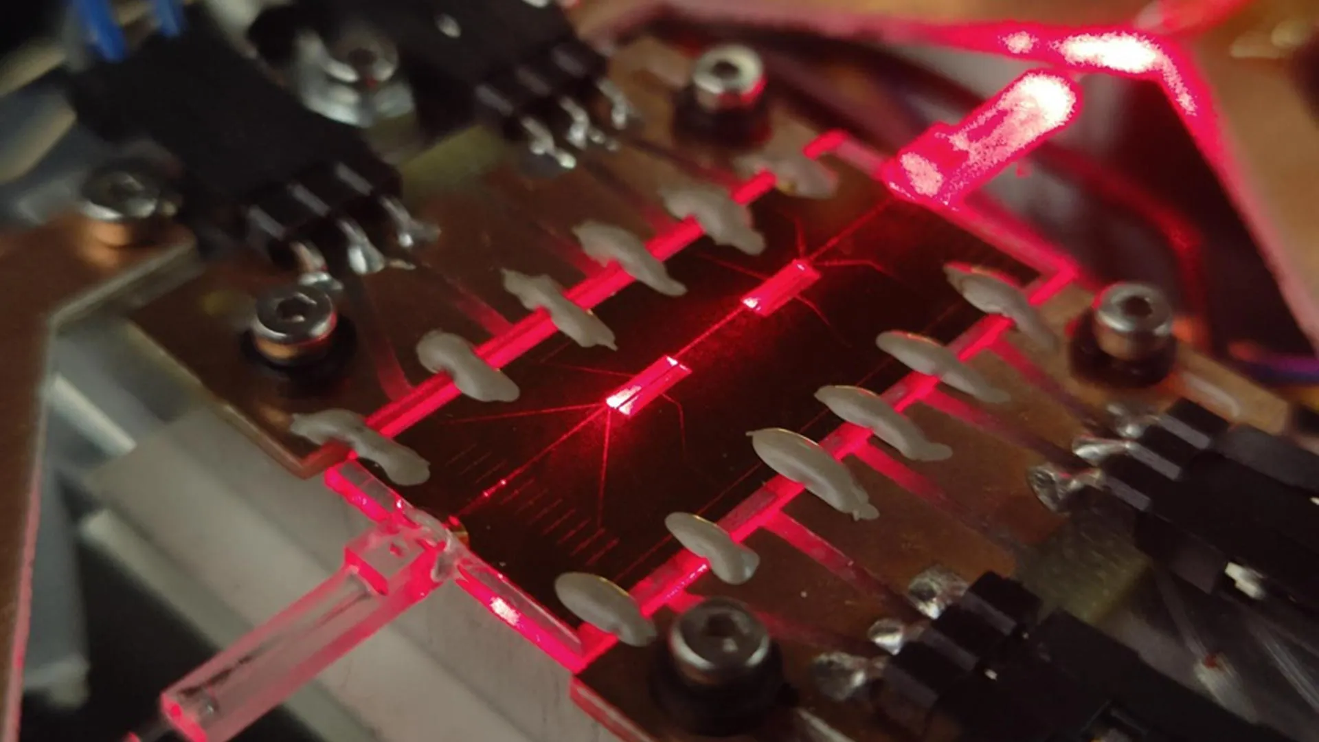
The operational ingenuity of the MOTE lies in its sophisticated use of light for both power and data transmission, circumventing the need for physical tethers or bulky internal batteries. The system leverages red and infrared laser beams, frequencies chosen specifically for their ability to safely penetrate brain tissue with minimal absorption and scattering. This external light source provides the necessary energy to power the minuscule device. Once energized, the MOTE then autonomously translates the detected electrical signals from neural activity into a series of infrared light pulses. This optical communication strategy is a key differentiator, offering a high-bandwidth, low-power method for data egress. The core of this system is a specialized semiconductor diode fabricated from aluminum gallium arsenide (AlGaAs). This particular material was selected for its dual functionality: it efficiently converts incoming light into electrical energy to power the internal circuitry, and it simultaneously acts as an emitter, generating the infrared light pulses that carry the encoded brain data back out. Integrated within this minute package are a low-noise amplifier, crucial for boosting the faint electrical signals from neurons while minimizing interference, and an optical encoder, responsible for converting these amplified electrical signals into the precisely timed light pulses. Both these components benefit from the same advanced semiconductor technology routinely employed in contemporary microchips, showcasing the translation of established silicon valley fabrication techniques to bio-integrated systems.
A particularly innovative aspect of the MOTE’s communication protocol is its reliance on pulse position modulation (PPM). This advanced encoding scheme, widely utilized in high-efficiency data transfer applications such as satellite communications and fiber optics, is exceptionally power-efficient. Rather than varying the amplitude or frequency of the light pulses, PPM encodes information by shifting the timing of fixed-amplitude pulses. This method allows for the transmission of significant amounts of data using minimal energy, which is paramount for a device operating without an internal power source. The MOTE’s ability to successfully transmit high-fidelity neural data wirelessly for over a year underscores the robustness and efficiency of this optical power and communication paradigm. This long-term functionality is transformative for longitudinal studies, enabling researchers to track neural changes over months, rather than days or weeks, offering unprecedented insights into chronic conditions and developmental processes.
The implications of this microscale neuro-implant are far-reaching, promising to revolutionize several domains within neuroscience and beyond. For fundamental brain research, the MOTE facilitates entirely new experimental designs. Scientists can now monitor the neural activity of freely moving animals for extended durations, observing natural behaviors, learning processes, and social interactions without the artifacts introduced by physical tethers or the stress of frequent handling for battery replacement. This capability will significantly enhance our understanding of complex brain functions, including memory formation, decision-making, and the neurological underpinnings of various psychiatric and neurological disorders. The unparalleled duration of monitoring means researchers can track the progression of diseases like Alzheimer’s or Parkinson’s in real-time within the same subject, observing the subtle neural changes that precede overt symptoms or monitoring the long-term efficacy of new therapeutic interventions.
Beyond basic research, the clinical potential of this technology is immense. One critical advantage of the MOTE’s design, specifically its material composition, is its potential compatibility with Magnetic Resonance Imaging (MRI) scans. Current neural implants often contain metallic components that can distort MRI images, create heating artifacts, and even pose safety risks within the powerful magnetic fields of an MRI scanner, thereby limiting the concurrent use of these two crucial diagnostic tools. The MOTE’s construction, primarily utilizing non-ferromagnetic semiconductors like aluminum gallium arsenide, suggests a pathway toward implants that can operate safely and transparently during MRI examinations. This would be a game-changer for diagnostics, allowing clinicians to simultaneously record neural activity and visualize brain structures or pathologies with high resolution, leading to more precise diagnoses and treatment planning for conditions such as epilepsy, brain tumors, or traumatic brain injury. Imagine the ability to precisely localize seizure onset zones through real-time neural recording while simultaneously mapping brain anatomy.
Furthermore, the adaptability of this microscale technology extends beyond the cerebral cortex. Researchers envision its application to other critical areas of the central and peripheral nervous system, including the spinal cord. Spinal cord injuries, for instance, could benefit from long-term monitoring of neural regeneration or the efficacy of rehabilitative therapies. Similarly, monitoring nerve activity in peripheral nerves could provide new insights into chronic pain conditions or facilitate more intuitive control of advanced prosthetics. The ability to integrate such unobtrusive sensors throughout the body could pave the way for a new generation of bio-integrated devices capable of continuous, real-time physiological monitoring, transcending the current limitations of wearable external devices.
Looking further into the future, the MOTE technology could become a foundational component for advanced neuro-prosthetics and integrated medical systems. The concept of embedding opto-electronics directly into artificial skull plates, for example, represents a vision where monitoring and even therapeutic interventions could be seamlessly integrated into a patient’s cranium. Such ‘smart’ skull plates could house an array of these micro-implants, enabling pervasive, long-term brain activity recording and potentially closed-loop stimulation systems that respond dynamically to neurological states, all while being largely imperceptible and requiring minimal maintenance. This could lead to highly personalized treatments for neurological disorders, where devices adapt in real-time to an individual’s unique brain activity patterns.
However, as with any transformative technology, challenges and ethical considerations must be carefully addressed. Scaling up production of such intricate microdevices, ensuring their long-term biocompatibility without triggering adverse immune responses, and developing robust algorithms to process the vast amounts of data these implants could generate are significant engineering hurdles. Moreover, the increasing integration of brain-interfacing technologies raises important ethical questions concerning data privacy, security, and the potential societal impact of such intimate human-technology interfaces. A comprehensive framework for responsible development and deployment will be essential as these technologies mature.
In conclusion, this development represents a monumental stride in neuro-engineering, setting a new benchmark for miniaturization, power efficiency, and chronic operational capability in neural implants. By surmounting many of the limitations inherent in previous generations of brain interfaces, this microscale optoelectronic device promises to unlock unprecedented insights into the brain’s complexities, accelerate the development of advanced diagnostics and therapeutics, and fundamentally reshape our understanding and treatment of neurological conditions. It marks a pivotal moment, ushering in an era where unobtrusive, long-duration neuro-monitoring becomes a tangible reality, laying critical groundwork for a future where seamless integration with the nervous system is not just an ambition, but an achievable scientific and medical frontier.