A revolutionary medical imaging methodology, developed by a collaborative team of researchers at Caltech and the University of Southern California (USC), is poised to transform diagnostic capabilities by offering unprecedented three-dimensional, colorized visualizations of the human body’s internal landscape. This innovative approach simultaneously captures the intricate physical architecture of soft tissues and provides real-time functional insights into vascular activity, including blood flow dynamics. Already deployed in preliminary human studies across various anatomical regions, this technology holds significant promise for advancing early disease detection, enhancing treatment monitoring, and deepening our understanding of complex biological processes, particularly in areas such as breast cancer diagnostics, the longitudinal tracking of diabetic nerve damage, and novel investigations into brain function.
The foundational details of this significant engineering and medical achievement were recently disseminated through a peer-reviewed publication in Nature Biomedical Engineering, signaling a major leap forward in non-invasive diagnostic modalities.
The Limitations of Conventional Imaging Modalities
For decades, medical professionals have relied on a suite of imaging tools, each offering distinct advantages but also presenting inherent limitations that often necessitate a fragmented diagnostic approach. Understanding these shortcomings provides crucial context for appreciating the innovation behind the new technique.
Standard ultrasound, for instance, remains a cornerstone of medical imaging due to its cost-effectiveness, portability, and real-time capabilities. It operates by emitting high-frequency sound waves and interpreting their echoes to construct images. While excellent for visualizing gross anatomical structures and dynamic processes like fetal movement or cardiac function, its primary output is typically two-dimensional, offering a limited field of view and often struggling to differentiate subtle tissue characteristics. Crucially, it provides little direct information about the metabolic activity or specific composition of blood within vessels, which can be vital indicators of pathology.
Photoacoustic imaging, a distinct yet related technique, offers a complementary perspective. It functions by directing short pulses of laser light into tissue. Certain molecules, such as hemoglobin in blood, absorb this light and subsequently generate tiny sound waves (thermoelastic expansion). These sound waves are then detected by ultrasound transducers and converted into images. This method excels at visualizing blood vessels in "optical color," which can convey information about blood oxygenation levels and flow dynamics, providing valuable functional data. However, a significant drawback of photoacoustic imaging alone is its comparatively poor ability to delineate detailed surrounding tissue morphology, meaning it struggles to provide the comprehensive anatomical context often required for precise diagnosis.
Beyond these two, other established imaging techniques like computed tomography (CT) and magnetic resonance imaging (MRI) also involve their own sets of trade-offs. CT scans utilize X-rays and sophisticated computer processing to generate cross-sectional images of the body. While highly effective for bone and dense tissue visualization, and capable of showing contrast-enhanced vascular structures, CT exposes patients to ionizing radiation, raising concerns about cumulative exposure, especially for repeated monitoring. Furthermore, it frequently requires the administration of iodine-based contrast agents, which carry risks of allergic reactions or kidney complications.
Magnetic resonance imaging (MRI), conversely, employs powerful magnetic fields and radio waves to produce highly detailed images of soft tissues without ionizing radiation. It offers superior contrast for many soft tissue types compared to CT. However, MRI examinations are notoriously time-consuming, leading to patient discomfort and susceptibility to motion artifacts. They are also significantly more expensive than ultrasound, require specialized infrastructure, and are contraindicated for patients with certain metallic implants or pacemakers. Many MRI protocols also benefit from, or even necessitate, the use of gadolinium-based contrast agents, which have recently raised concerns regarding retention in body tissues.
The collective implication of these individual limitations is that clinicians frequently need to combine information from multiple imaging modalities, subjecting patients to sequential scans, varying costs, and sometimes repeated exposure to radiation or contrast agents, thereby complicating and prolonging the diagnostic pathway.
The Genesis of RUS-PAT: A Synergistic Approach
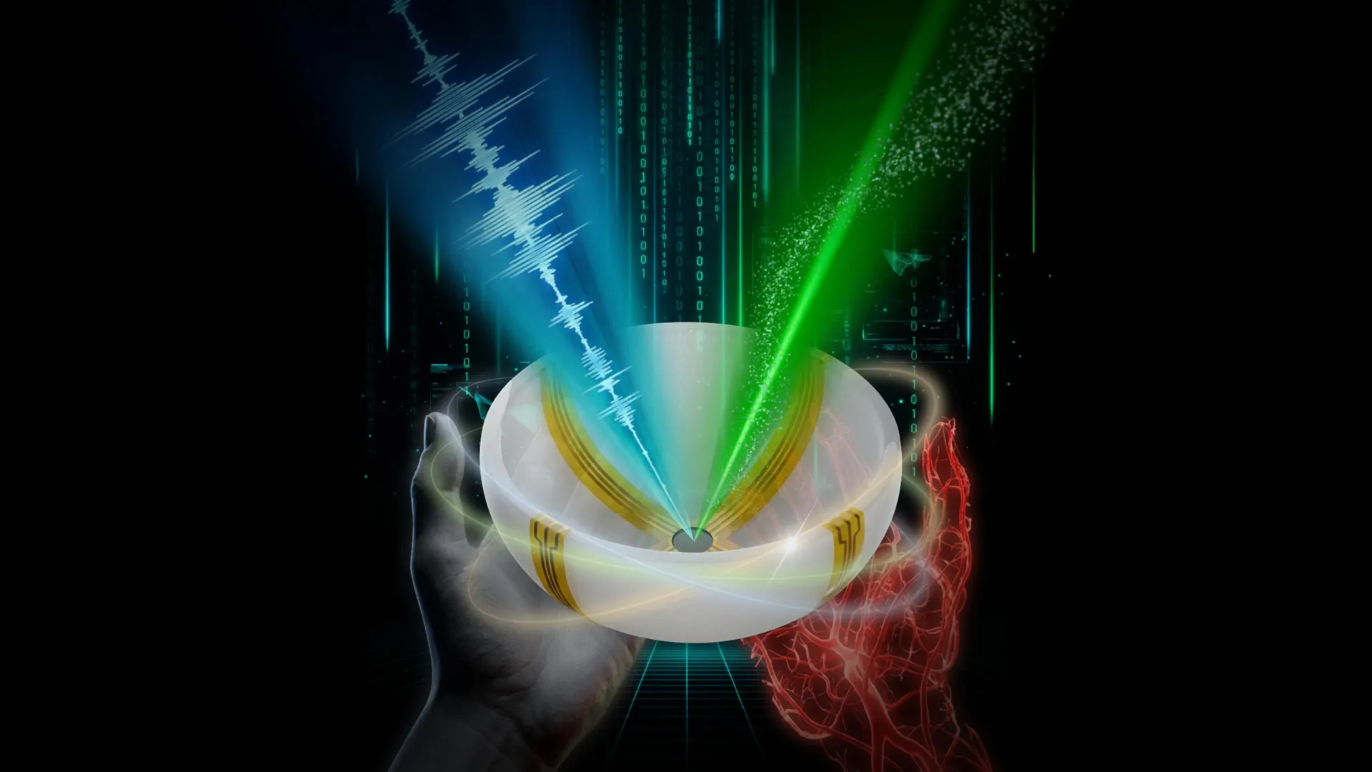
Recognizing the inherent strengths and weaknesses of existing technologies, the research team embarked on a mission to transcend these individual limitations by developing a novel hybrid system. The result is Rotational Ultrasound Tomography combined with Photoacoustic Tomography, or RUS-PAT. This sophisticated approach represents a strategic convergence of two powerful imaging principles, designed to leverage their complementary attributes.
The underlying photoacoustic tomography (PAT) component of RUS-PAT traces its lineage back over two decades to pioneering work by Lihong Wang, who serves as the Bren Professor of Medical Engineering and Electrical Engineering and the Andrew and Peggy Cherng Medical Engineering Leadership Chair at Caltech. In its fundamental operation, PAT capitalizes on the principle that when short laser pulses illuminate tissue, specific light-absorbing molecules within that tissue vibrate, generating transient acoustic signals. These sound waves are then detected, processed, and computationally reconstructed into detailed images that primarily highlight structures with strong light absorption, such as blood vessels.
Professor Wang, who also holds the position of Caltech’s executive officer for medical engineering, articulated the central objective of the RUS-PAT project: to seamlessly integrate the robust structural imaging capabilities of ultrasound with the rich functional and vascular information provided by photoacoustic imaging. He emphasized that this was not merely a simple additive process. "It’s not like one plus one," he explained, underscoring the complex engineering challenge involved in identifying an optimal methodology for truly merging these distinct physical modalities into a single, cohesive system that outperforms their individual sum. The goal was to create an integrated platform that could simultaneously deliver both detailed anatomical context and dynamic physiological insights within a single, rapid scan.
A Paradigm Shift in Design and Practicality
A critical hurdle in merging ultrasound and photoacoustic imaging lay in the inherent design complexities of traditional ultrasound systems. Conventional ultrasound typically employs an array of numerous transducers, each responsible for both emitting and receiving sound waves. Integrating such a multi-element system directly with photoacoustic imaging, which primarily requires only ultrasound detection, would result in an overly intricate and prohibitively expensive device, severely limiting its potential for widespread clinical adoption.
This disparity sparked a pivotal insight for Professor Wang. He recognized that photoacoustic imaging’s core requirement was the detection of ultrasound waves generated by light absorption, not the generation of ultrasound waves for structural imaging. This led to a novel conceptualization: "I thought, ‘Wait, can we just mimic light excitation of ultrasound waves in photoacoustic tomography, but do it ultrasonically?’"
This innovative thought process paved the way for a radically simplified and more practical design. Instead of relying on a multitude of transducers to actively generate ultrasound waves for anatomical imaging, the RUS-PAT system utilizes a single, wide-field ultrasound transducer. This transducer efficiently transmits sound waves throughout the targeted tissue volume. Crucially, the same set of detectors then captures the acoustic signals generated by both the initial wide-field ultrasound pulses (for structural imaging) and the photoacoustically generated sound waves from laser light absorption (for functional and vascular imaging).
The ultimate hardware configuration for RUS-PAT further embodies this minimalist yet highly effective philosophy. It employs a small number of strategically positioned arc-shaped detectors that are designed to rotate around a central axis. This rotational mechanism is key: by moving the detectors, the system effectively simulates the data acquisition capabilities of a much larger, full hemispheric detector array. This ingenious design significantly reduces the number of physical components required, thereby making the system far simpler to manufacture, less expensive to produce, and inherently more practical for eventual widespread clinical deployment, without compromising the quality or comprehensiveness of the acquired data.
Demonstrating Clinical Feasibility and Transformative Potential
The successful integration of acoustic and photoacoustic principles within the RUS-PAT framework has already yielded compelling evidence of its clinical utility. Dr. Charles Y. Liu, a co-author of the study and a visiting associate in biology and biological engineering at Caltech, highlighted the significance of these findings. Dr. Liu, who also holds positions as a professor at the Keck School of Medicine of USC, director of USC’s Neurorestoration Center, and chair of neurosurgery at the Rancho Los Amigos National Rehabilitation Center, stated, "The novel combination of acoustic and photoacoustic techniques addresses many of the key limitations of widely used medical-imaging techniques in current clinical practice, and, importantly, the feasibility for human application has been demonstrated here in multiple contexts."
Because the RUS-PAT method operates wherever light can penetrate tissue, either directly or via endoscopic delivery, its potential clinical applications are remarkably broad.
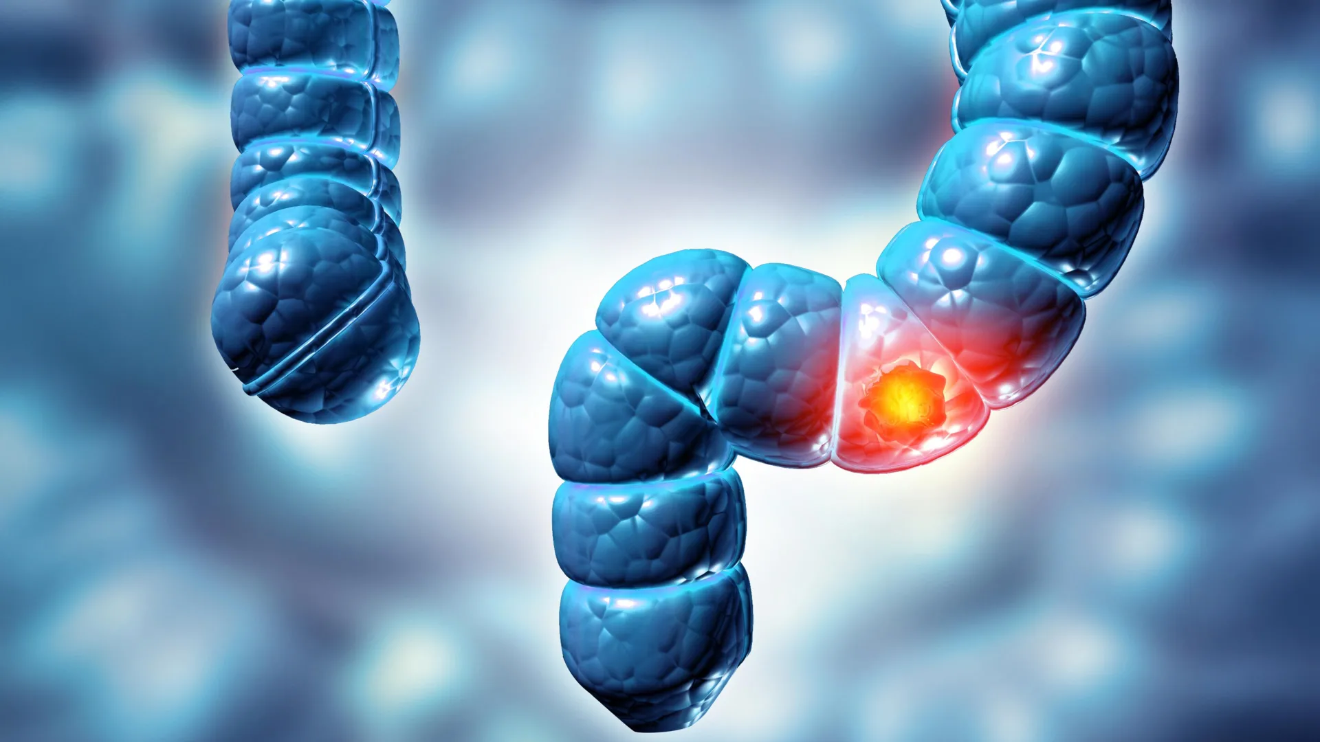
In the realm of breast cancer imaging, RUS-PAT could usher in a new era of diagnostic precision. By simultaneously providing detailed anatomical location of a suspicious mass (structural information) and revealing critical biological activity within it, such as neo-angiogenesis (formation of new blood vessels characteristic of tumors) and altered oxygenation (functional information), clinicians could gain a more comprehensive understanding of a lesion’s nature. This dual insight could potentially improve the accuracy of early detection, better differentiate benign from malignant lesions, guide biopsy procedures with greater precision, and more effectively monitor treatment response, potentially reducing the need for invasive procedures or repeated imaging.
For patients afflicted with diabetic neuropathy, a debilitating complication of diabetes affecting nerve function, RUS-PAT offers a unique diagnostic advantage. Current monitoring often involves separate assessments of nerve structure and vascular health. This new technique could enable physicians to simultaneously visualize the delicate structure of peripheral nerves, identifying any signs of degeneration or damage, while also assessing the local oxygen supply and microvascular integrity in a single, non-invasive scan. This integrated perspective is crucial for tracking disease progression, evaluating the efficacy of interventions, and potentially predicting areas at higher risk for complications, thereby improving patient management and quality of life.
Professor Wang also underscored the profound implications for brain research. While current brain imaging technologies are powerful, simultaneously observing fine brain anatomy and dynamic blood flow changes, which are often proxies for neuronal activity, remains challenging, especially for superficial cortical layers or through minimally invasive approaches. RUS-PAT could provide an unprecedented window into the living brain, allowing scientists to study intricate anatomical details alongside real-time blood flow dynamics. This could accelerate discoveries in understanding neurological disorders, deciphering cognitive processes, and developing advanced brain-computer interfaces, particularly when combined with endoscopic light delivery for deeper access.
Operational Efficiency and Future Directions
The RUS-PAT system boasts impressive operational metrics that underscore its clinical viability. Each scan can be completed in less than one minute, a significant advantage over many existing modalities. This speed not only enhances patient comfort but also improves clinical throughput, allowing more patients to be examined efficiently.
Currently, the system is capable of imaging tissue up to approximately 4 centimeters deep. While this depth is suitable for numerous superficial applications (e.g., skin, breast, peripheral nerves), the research team is actively exploring methods to extend its reach. One promising avenue involves the use of endoscopic tools to deliver laser light and collect signals from deeper within the body, thereby expanding the technique’s applicability to internal organs and structures.
The existing setup integrates ultrasound transducers and a laser beneath a scanning bed, optimized for patient comfort and efficient data acquisition. Crucially, the technology has already moved beyond theoretical models and laboratory prototypes, having undergone successful testing on human volunteers and patients. This critical step signifies its readiness for real-world application and marks its entry into the early stages of the rigorous pathway toward broader clinical integration and widespread medical use.
Collaborative Research and Funding Support
The seminal paper detailing this breakthrough, titled "Rotational ultrasound and photoacoustic tomography of the human body," represents a significant collaborative effort. The co-lead authors of the publication are Yang Zhang, Shuai Na, and Dr. Jonathan J. Russin. Zhang and Na conducted their pivotal research as postdoctoral researchers at Caltech and have since transitioned to academic positions at Tsinghua University and Peking University in Beijing, respectively. Dr. Russin is affiliated with the Keck School of Medicine of USC and the Rancho Los Amigos National Rehabilitation Center in Downey, California, bringing critical clinical expertise to the project.
Additional contributors from Caltech include Karteekeya Sastry, Li Lin, Junfu Zheng, Yilin Luo, Xin Tong, Yujin An, Peng Hu, and former research scientist Konstantin Maslov. Dr. Lin is currently based at Zhejiang University in Hangzhou, China. Dr. Tze-Woei Tan from the Keck School of Medicine of USC also served as a co-author. This extensive interdisciplinary collaboration highlights the complex nature of such advanced biomedical engineering endeavors. The research received vital financial backing from the National Institutes of Health, underscoring the federal commitment to fostering innovative medical technologies that promise to revolutionize healthcare.
The advent of RUS-PAT heralds a transformative era in medical diagnostics and research. By seamlessly integrating structural and functional imaging in a non-invasive, rapid, and cost-effective manner, this technology offers a powerful new lens through which to observe the complexities of human health and disease, ultimately paving the way for more precise diagnoses, personalized treatments, and deeper scientific understanding.